Typical Plant Cell Microscope Slides Typical plant cell, Plant cell, Plant and
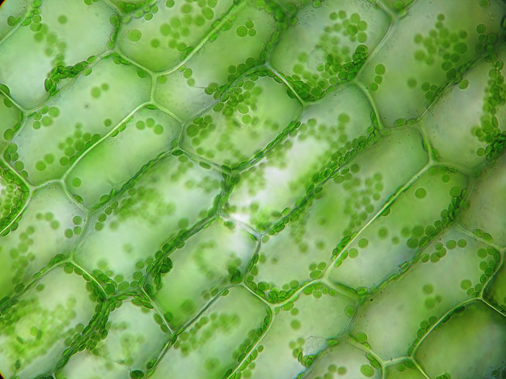
What you see when looking at an elodea leaf under a microscope.
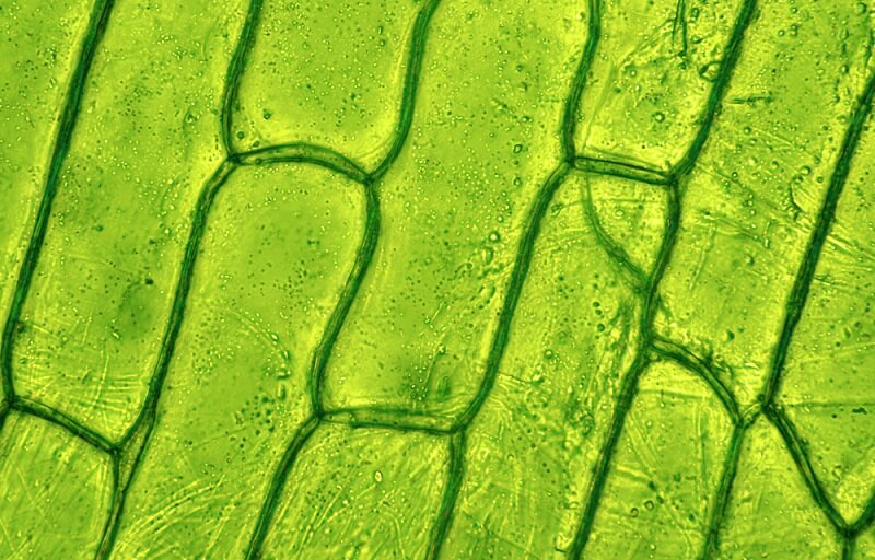
plant cell microscope images Biological Science Picture Directory
What Are the Differences Between a Plant & an Animal Cell Under a Microscope? ••• Updated May 14, 2019 By Jack Powell All living things are made up of cells. Some of the smallest organisms, such as yeast and bacteria, are single-celled organisms, but most plants and animals are multicellular.

Chloroplasts in Elodea cells, light micrograph Stock Image C038/6969 Science Photo Library
It is used primarily as a structural component in plant cell walls. Chloroplasts are possibly the most noticeable organelles in plant cells.. Use two hands to carry the microscope. Place one hand under it to support its weight, and hold onto the handle on the back of the microscope arm. If your microscope does not have a handle, hold tightly.

Phloem Plant Cells Photograph by Dr Keith Wheeler/science Photo Library Fine Art America
Overview of a flowing plant The Roots The Stem - Xylem and Phloem The Leaves The Flowers The Seeds Why microscope is important in biology? The microscope is a very important tool in a biological laboratory. Many cellular structures are too tiny to see by naked eyes.

Plant cells microscopy
While there are many forms of microscopy, this activity provides guidance and advice on sample preparation for a brightfield microscope, along with safe and easy-to-use stains like toluidine blue to visualize and identify plant parts (Figure 1).
Plant Cell Under Microscope 40X Labeled 1 Chloroplast and cell wall animal cell
Plant cells under the microscope - YouTube 0:00 / 2:50 Plant cells under the microscope Science Skool 4.75K subscribers Subscribe Subscribed 143 30K views 5 years ago A short video showing.

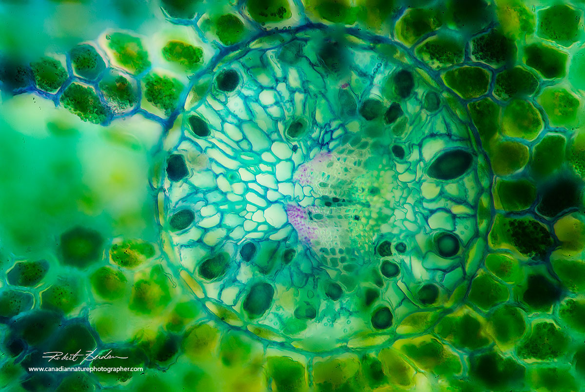
Cross section of a plant stem under a microscope. Biology art, Plant cell, Bio art
It was not until good light microscopes became available in the early part of the nineteenth century that all plant and animal tissues were discovered to be aggregates of individual cells. This discovery, proposed as the by Schleiden and Schwann in 1838, marks the formal birth of cell biology.
Modeling a Plant Cell Perkins eLearning
Allow the nail polish about four hours to dry. Using a pair of tweezers, peel off a film (thin skin) from the surface of the leaf. Gently place the film onto a microscope slide and cover with a cover slip. Start with low power and increase to 100x (frequency of stoma can be counted at 100x) Record your observations.

Plant Cell Under Microscope Labeled Pin By Nia On Education Plant Cell Electron Microscope
Witness a living plant cell's chromosomes carrying genetic material duplicate during the process of mitosis. Time-lapse photography of a live plant cell nucleus undergoing mitosis. Examine the structures adenine, ribose, and a three-phosphate chain in adenosine triphosphate molecule and their role in releasing energy for cellular activities.
Plant cells under the microscope r/MicroPorn
View under the microscope using the highest magnification for the best cellular details and draw what you see. Be sure to indicate the magnification used and specimen name. Also, indicate the estimated cell size in micrometers under your drawing. Figure 7.. What are the distinguishing characteristics of a plant cell versus an animal cell?
The Microscopic Beauty of Plants and Trees by Robert Berdan The Canadian Nature Photographer
plant cell, the basic unit of all plants. Plant cells, like animal cells, are eukaryotic, meaning they have a membrane-bound nucleus and organelles. The following is a brief survey of some of the major characteristics of plant cells. For a more in-depth discussion of cells, see cell. Unlike animal cells, plant cells have a cell wall surrounding.
Plant cells under the microscope. pics
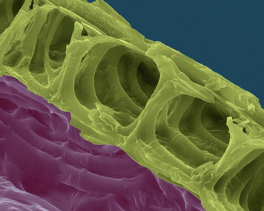
Figure 10.1.5 10.1. 5: A micrograph of a cell nucleus. The nucleolus (A) is a condensed region within the nucleus (B) where ribosomes are synthesized. The nucleus is surrounded by the nuclear envelope (C). Just oustide the nucleus, the rough endoplasmic reticulum (D) is composed of many layers of folded membrane.
Plant Cell Wall Microscope Image Micropedia
Step by step Click to see a step-by-step slideshow. YOU WILL NEED: An onion, a slide and cover slip, a cotton bud, some food colouring, a plate to put the cotton bud on and of course a.

Plant Cell Under Microscope
Light microscope micrograph showing mitosis in onion cells of the root meristem. At bottom, is a cell in anaphase. On the top right corner, a prophase. Cross-section Plant Stem under the microscope for classroom. of 100. Search from 6,839 Plant Cell Microscope stock photos, pictures and royalty-free images from iStock.

Botany Professor Everything you wanted to know about plant cells, but were afraid to ask
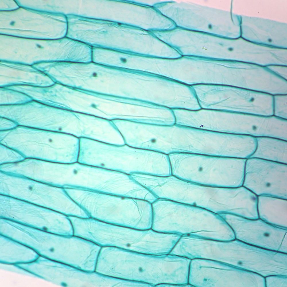
Purpose: Students will observe plant cells using a light microscope. Two cells will be observed, one from the skin of an onion, and the other from a common aquarium water plant (anacharis).. View under the microscope and sketch the cells at each magnification. Label the cell wall, nucleus, and cytoplasm as they appear under high power..
Eukaryotic Cell The Definitive Guide Biology Dictionary
Putting plants under the microscope Words: Kathy Grube Layout: Jacqueline Garget Published: 18 June, 2021 Biosensor imaging of a seedling, measuring how the concentrations of the plant hormone gibberellin change as the plant grows. Credit: Annalisa Rizza. Humans have been making use of plants for thousands of years.
